The standalone DHM®-T evolutive solution
A complete solution from live cell environment to reporting
Lyncée delivers turn-key systems that include:
- a modular and evolutive inverted DHM® T microscope
- sample environmental control
- multi-modal imaging add-ons
- motorized samples stage for automation and screening
- software with automated workflows, data analysis and reporting
The modular and evolutive DHM®-T fits any budget
The DHM® T-100 is the simplest of Lyncée inverted microscope offering an ideal cost effective tool for routine and non-perturbing cell culture monitoring at single and population level. It is equipped with a single fixed objective, that can be selected from large choice of magnification, numerical aperture, and coverslide corrections.
The DHM® T-100 can then later by upgraded to a DHM® T-1000 that support multiple objectives operation.
Optional accessories for living cells handling and workflow control
Lyncée options range is fully compatible with any DHM® of the T series. The principal ones are the:
- motorized stage enabling automation of multi-site measurement, including a large variety of samples holders
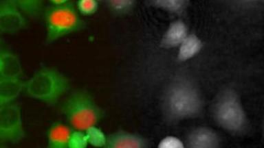
- fluorescence module, used in conjunction with Transmission DHM® the fluorescence module by Lyncée Tec provides simultaneous real time quantitative phase measurements and videos of epifluorescence characterization on a single platform, through the same objective lens. It enables to compare High Content Screening DHM® measurements with reference fluorescence assays, or for single cell analysis after segmentation. It includes standard and high end fluorescence cameras and light sources
- Rescanning confocal module: three modalities in a single instrument: QPM by DHM®, Epifluorescence, and confocal scanning.
- environmental chamber, enabling long duration time lapse of living cells without perturbing them
Software for acquisition, data analysis, interpretation, and reporting
The proprietary software Koala Acquisition & Analysis controls the measurement acquisition parameters, controls the laser source, the electronic and peripheral equipment such as the motorized stage.
Cell Analysis Tool automatically processes the acquired hologram or reconstructed phase images, refocus the cells after the acquisition (a unique feature of holographic microscopy), track the spatial displacement of cell during time-lapse experiments, and extracts the biological information for the Single site and Multiple site workflows, and combine measurements of Multi-modal experiments. It save each analysis automatically in a XLSX file, and produce a PDF report summarizing the key feature of the analysis.