DHM®: a simple workflow
From measurement to experiment reporting
Lyncée Tec provides a whole solution for the entire experimental workflow, from sample preparation, to image acquisition, data processing and reporting.
Both DHM® T series and DHM® cameras are fully compatible with this workflow.
You can thus spend more time designing experiments and less time doing and analyzing them.
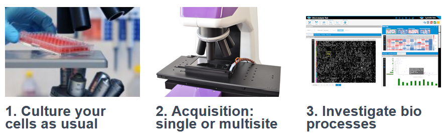
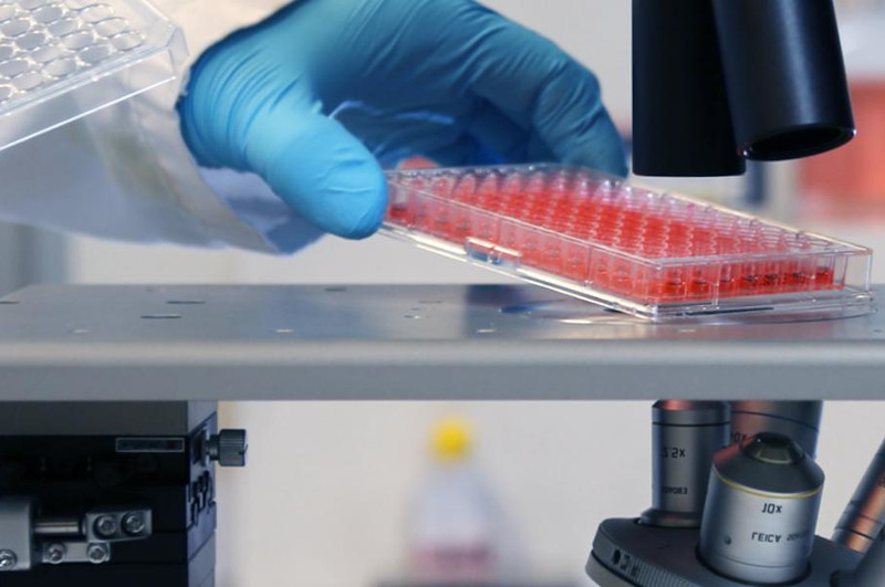
1. Culture your cells as usual
Culture you sample as usual on any transparent medium: microscope slides, petri dishes, multiwells plates, in perfusion chambers or microfluidic devices.
No needs of harmful marker, no pipetting steps which can detach your cells. DHM® is a truly non-perturbing system. You are immediately ready to go!
NO NEEDS of:
- specific substrate
- staining cells
- pipetting and washing step
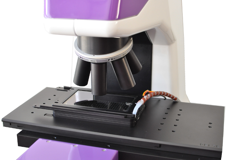
2. Acquisition: single and multi-site time-lapses
Fast and long time-duration time-lapses
DHM® image your cells at up to 194 fps with a standard system, and up to 100’000 fps an with the optional ultra-high speed cameras. As each image is captured in less than one millisecond, fast dynamical processes can be investigated.
Using our environmental control chamber, and as DHM® measure you can monitor your cells over multiple days without perturbing them.
Multiple-sites measurements
With our motorized stages, extend the field of view by stitching multiple images together, and/or monitor your cells at multiple site, for performing for instance multi-well plates screening.
Of course, both time-lapses and multiple sites monitoring capabilities can be combined for advanced workflows.
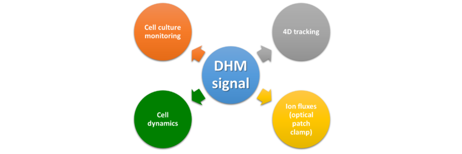
3. Investigate bio processes
Lyncée Tec’ intuitive CAT software, specifically designed for life sciences experiments, provides automatically data, plots, and reports of your experiment. It supports single site (individual cells and population analysis), multi-sites (end-points, dose response), time-lapse, multi-modal (correlative microscopy) and 4D-tracking workflows.
PDF reports summarizing the key feature of the analysis together with the relevant plots can be exported at the end of the analysis to keep precise track of your experimental work.
Data are available for advanced analysis in understandable formats compatible with Excel, ImageJ, and most of the software used in cellular imaging laboratories.