Correlative microscopy
Spatial and temporal correlation
Open new perspectives with multi-modal acquisitions with our DHM® microscopes or cameras.
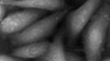
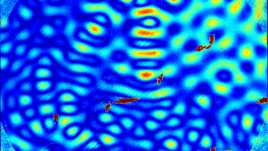
By combining Quantitative Phase Imaging (QPI) and fluorescence you obtain better insights into your monitored biological processes.
- Quantitative Phase Imaging (QPI) offers label-free measurements of cell morphology and dry mass, two unique biomarkers for the cell’s physiological state
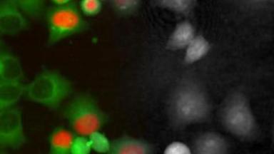
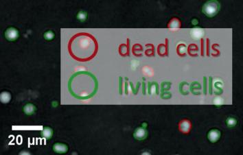
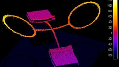
- Fluorescence adds the specificity associated with the labeling of targeted cellular components or molecules
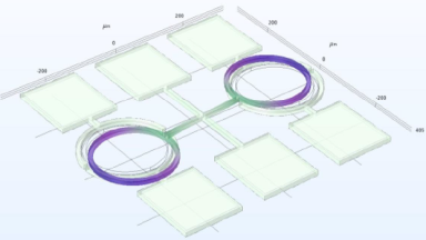
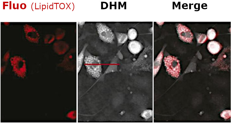
Spatial co-localization
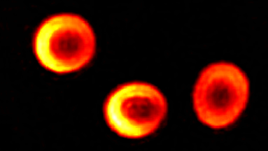
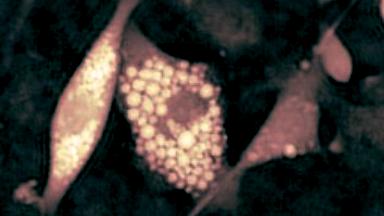
Spatial co-localization thanks advantage of the specificity of fluorescence to identify distinct cellular structures.
Find more about how this spatial co-localization can be used to identify lipid droplets here.
Temporal co-localization
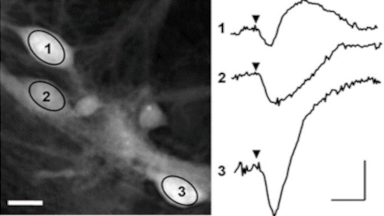
Temporal correlation between QPI and fluorescence provides valuable information on the dynamics of the monitored processes regarding the synchronization of the bio-processes.
Find more about how this temporal correlation can be used to investigate cardiomyocytes beating here.