Cardiomyocytes
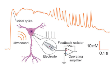
Cardiomyocyte beating monitoring
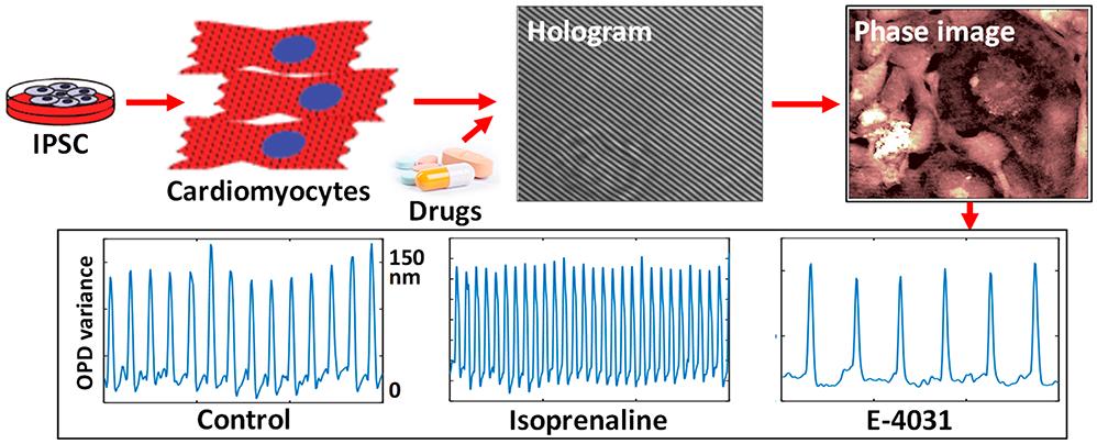
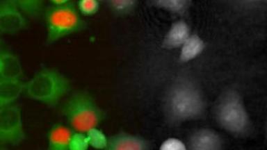
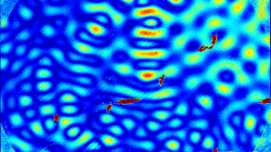
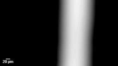
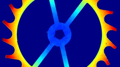
Cardiotoxicity is a major health issue and principal concern when developing novel therapeutic drugs. Combining Quantitative Phase Microscopy (QPM) and fluorescence allows to combine two different information for a better understanding of the physiological contraction of the cells.
Unique advantages of Lyncée Tec DHM®
- Non-perturbing measurement of the cardiomyocytes (no adverse effect due to fluorescence staining)
- Fast image acquisition (up to 194 fps)
- Access to temporal correlation with fluorescence
Material and methods
- Biological Model: iCell cardiomyocytes from Cellular Dynamics (Fujifilm)
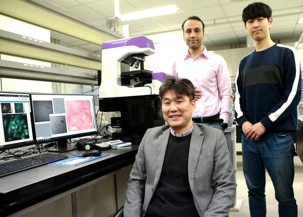
- DHM®: T1000, 20x/0.4 NA with fluorescence module, and environmental control chamber.
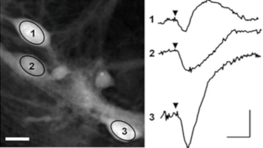
- Data analysis: automatic dual recording of phase and fluorescence (Fluo-4) modalities from the same field of view
- 14 parameters of the beating profile are measured to quantify its dynamics
Results
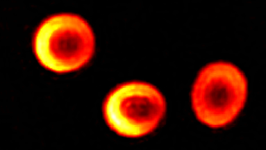
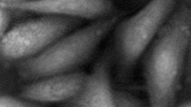
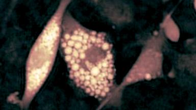
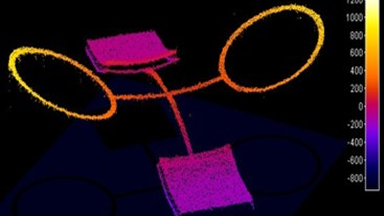
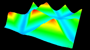
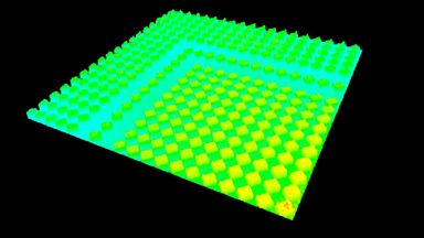
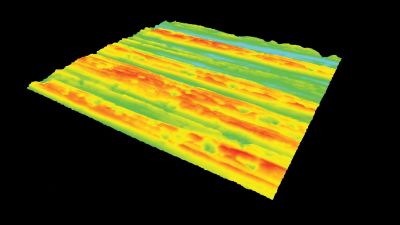
Dry mass movements recorded by DHM provide mechanical (contraction/relaxation) information and Fluo-4 fluorescent dye provide intracellular calcium release information. Both are temporally correlated to provide insights on the beating dynamics, as shown by the early pic of calcium which triggers the mechanical contraction of the cells.
Publication
Marker-Free Automatic Quantification of Drug-Treated Cardiomyocytes with Digital Holographic Imaging
K. Jaferzadeh et al., ACS Photonics. 2020, 7, 1, 105-113
Our QPI-fluorescence multimodal microscope purchased at Lyncée Tec is a key addition to our list of equipment to study cellular dynamics. It allows us to combine simultaneous biomechanical data obtained with QPI with changes of ions fluxes recorded by fluorescence for high-content screening applications.
With this unique system and its analysis software, we have access to a comprehensive understanding of cell structure and dynamics with nanoscale sensitivity. For instance, we recently use it to obtain a full characterization of the physiological effect of cardiac drug candidates.
Lyncée Tec microscope greatly increases our throughput and opens new research perspectives.