Quantitative phase imaging of living cells
Phenotyping by DHM®
Marker free, non perturbing Quantitative Phase Imaging (QPI)
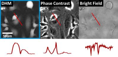
Cells are so-called “phase objects”. Without the use of any marker, they modify the phase of the light which cross them, but have almost no effect on its intensity. Differential Interference Contrast (DIC) and other phase contrast imaging methods exploit this property, generate a contrast from transparent cells, and enables to visualize them. But they do not measure quantitatively the phase delay of the wave crossing cells.
The transmission Digital Holographic Microscope and Cameras from Lyncée Tec, based on the DHM® technology, provide quantitative phase images that are directly proportional to the morphology and the intracellular protein contents (dry mass) of the cells.
These two recorded parameters are key endogeneous biomarkers. They are measured with non-focused and low intensity illumination, preventing cells from photo-toxicity and photo-bleaching. With DHM®, your cells are measure without any perturbation. Very long duration time-lapse protocol can be performed, and cells responses will be uniquely due to their biological environment and to the experimental protocol under investigation. (more about it)
Morphology and intracellular content: the key endogenous biomarkers
The DHM® technology takes advantage of the natural endogenous contrast of cells: morphology and intracellular content. There is no need to use interfering external dyes.
Indeed, these biomarkers have not only an interest in terms of biomarkers. They have additionally an inestimable value to monitor cell heath and underlying physiopathological processes. For more than 20 years, Lyncée Tec has closely cooperated with several pioneer research labs, to investigate and demonstrate it. Resulting in numerous seminal publications establishing the wealth of QPI by DHM®.
Our CAT software for bioanalysis has been matured continuous during this fundamental scientific work. It allows first to easily extract all the rich information contained in the quantitative images in term of morphology, intracellular content, dynamic parameters, and population metrics, and second to use them for many biological paradigms, as illustrated by examples in the examples of the next paragraph.
DHM® monitors cell health state and underlying physiopathological processes
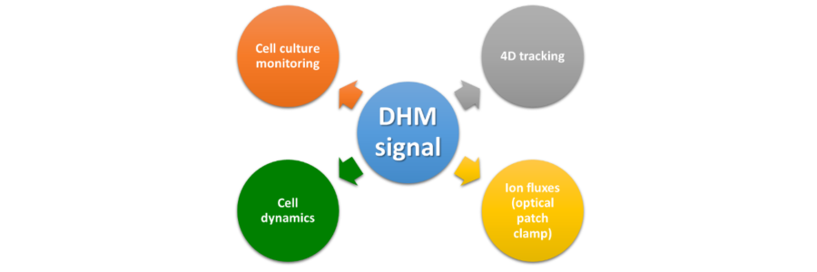
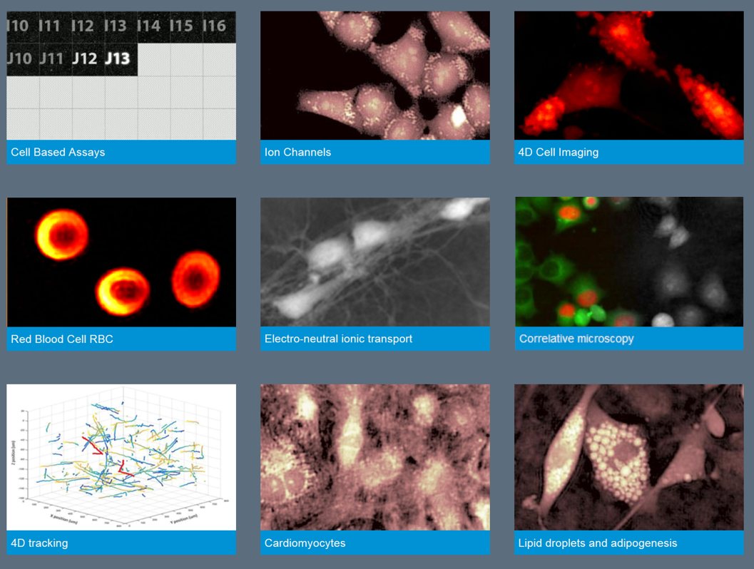
Here below are a few application examples of living cells quantitative phase imaging by DHM® and measurement interpretation:
- Cytotoxicity
- Cell migration/proliferation
- Cardiomyocytes dynamics
- Hematology
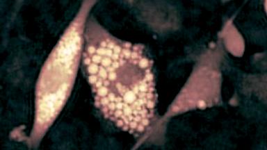
- Adipogenesis
- 4D tracking
- Drug screening
- Optical patch clamp
- Fluorescence correlation
Competitive advantages of DHM
DHM® offers key advantages when live samples need to be recorded in a non-perturbing way.
- Speed: microsecond acquisition
- Label-free: no marker needed, no phototoxicity (unlike fluorescence)
- Non-invasive: non-perturbing physiological measurements
- Quantitative: recordings proportional to physiological parameters
- High content analysis: large set of parameters acquired
- Perfect-focus: Off-line refocusing capability
- Ease of use: no sample preparation, direct imaging (no focusing or scanning)
These advantages combined allow to monitor living cells in real time for an extended period of time. The absence of label allows for repeated measurement and even to perform an invasive measurement at the end of the DHM recording using another modality (for instance, fixation and antibody labeling).
Multimodality
The high content information provided by DHM can be correlated with simultaneous fluorescence measurements of the same field of view with the addition of the optional fluorescence module.